Solving Diabetes Retinopathy Problems
Whether you have diabetes or not, problems with the eyes are inevitable as you age. But if you have diabetes, you are likely to experience more eye problems and at an earlier age than people without diabetes.
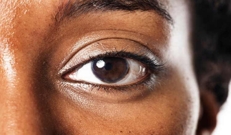
Visual images, or the things you see in the world around you, come into your eye as light. Light enters the eyes through your pupil. The cornea and lens that cover your pupil focus the light, which then travels through the posterior segment of your eye. This part of your eye is filled with a gel-like substance known as the vitreous humor. The light passes through the vitreous humor to the back of the eyeball until it hits your retina, the light-sensing part of your eye.
Light hitting on the retina transmits signals to your brain through the optic nerve. These signals allow you to see. The brain processes these signals as visual images, or sight. Other nerves also control the way your eyes move, how they focus, and how they adjust pupil size to allow for changes in lightness and darkness.
For your eyes to function properly, you need a good, steady supply of blood to the retinas, to bring them the nutrients they need. Damage to these blood vessels can threaten your vision. Your eyes also depend on healthy nerves to transmit optical signals and to control eye movements. If any of these nerves are damaged, you will experience vision problems. Because diabetes can cause damage to both blood vessels and nerve fibers, the eyes are especially susceptible to problems in people with diabetes.

Some of the eye problems causes by diabetes are mild and easily reversed. Often, maintaining good blood glucose control is enough to make them go away. But other problems can jeopardize our sight and cause blindness if not treated properly. Almost all vision problems triggered by diabetes are treatable if caught early enough. However, most vision problems are not painful and may at first seem like minor nuisances. It is easy to put off doing something about these problems.
But that’s the worst thing you can do. Anytime you notice a change in how you see things, contact your eye doctor right away. And even if you don’t notice any changes, you should see your eye doctor at least once a year. You may even be completely unaware of some of the changes caused to your eyes by diabetes. Therefore, it is extremely important that you see an eye-care specialist on a regular basis.
DIABETES RETINOPATHY
The retina is the light-sensing part of your eye, located along the back wall of your eye. It contains the rod and cone cells, which detect light and send a signal to your brain. The retina contains many small blood vessels that supply the retina with blood, oxygen and nutrient it needs to function. Unfortunately, these blood vessels can be easily damaged. Poor blood glucose control can change the way your blood flows and can weaken the walls of these blood vessels. When the blood vessels become damaged, retinopathy can occur.
Early on, damage to the blood vessels is minimal and can be easily treated. This stage is known as non-proliferative retinopathy. If left untreated, it can progress to proliferative retinopathy, a much more dangerous condition. During either stages of retinopathy, macular edema can occur. This is caused by leakage of fluid into the macula, the region of the retina responsible for color vision and visual acuity. Macular edema can cause loss of vision if not treated.
If you have type 1 diabetes, you will probably not develop retinopathy until you have had diabetes for several years. After 10 years, however, more than half of people with type 1 have retinopathy in one form or another. After 15 years, almost all people with type 1 diabetes have retinopathy. However, only half of all people with type 1 diabetes will develop proliferative retinopathy.
On the other hand, if you have type 2 diabetes, you are more likely to have retinopathy at diagnosis and more likely to develop retinopathy early on in the course of diabetes. Twenty percent of all people with type 2 diabetes will have retinopathy diagnosis. After 15 years, 60% to 85% of people with type 2 diabetes have retinopathy. At this point, up to 20% will also have proliferative retinopathy.

The good news is that retinopathy is preventable. The Diabetes Control and Complications Trial (DCCT) showed that maintaining tight control of blood glucose levels reduced the risk of retinopathy by 76%. Early treatment and maintaining good blood glucose control can go a long way in preventing vision loss from retinopathy.
DIABETES NONPROLIFERATIVE RETINOPATHY
If you maintain poor control of blood glucose, the blood vessels that supply your retina can become damaged. When this happens, the blood vessel walls can balloon out, creating tiny aneurysms. This causes the tissue in your retina to swell, a condition known as edema. The blood vessels can also rupture, or hemorrhage, and leak fluid or blood into the retina. When this occurs, you have nonproliferative retinopathy.
Your retina is cut off from the supply of oxygen and nutrients it needs and doesn’t function properly. At this stage, you may experience vision problems or you may be unaware of the problem. If detected early enough, nonproliferative retinopathy can be treated and reversed. However, if left untreated, it can progress to proliferative retinopathy.
DIABETES NONPROLIFERATIVE RETINOPATHY SYMPTOMS
Unfortunately, nonproliferative retinopathy is often symptomless, especially in the early stages. You may find that parts of your field of vision are distorted, but in the early stages you will probably not experience vision loss, unless macular edema is also present.
Although you may not feel symptoms, your eye doctor can detect nonproliferative retinopathy. The only way to know for sure is through a thorough eye exam conducted by an ophthalmologist.
What You Should Do
If you have diabetes, you should have a yearly eye exam by an ophthalmologist whether or not you have any symptoms. If you have any symptoms or notice any changes in your vision, tell your doctor or ophthalmologist right away. During your eye exam, your doctor should measure how well you can see (visual activity), evaluate how you move your eyes, determine whether you need glasses, screen for glaucoma and cataracts, and evaluate changes in both color perception and night vision. Your retina should also be thoroughly examined. This is done by first dilating the pupil, then using one of several techniques to examine the retina. Using an ophthalmoloscope, your ophthalmologist can get a magnified view of your retina and look for any changes or irregularities of the optic nerve, the macula and the blood vessels of the retina. If there is any hemorrhaging, swelling or new blood vessel formation, your ophthalmologist can detect it.

Your ophthalmologist may also perform a fluorescein angiography test. Using this method, a fluorescent dye is injected into your arm. The flow of dye through the retina is tracked using special camera. Healthy blood vessels will keep the blood flowing and will not leak, but damaged blood vessels will leak blood and dye. This will help your doctor pinpoint areas in your retina that may require treatment. It will also reveal whether you have any macular swelling that could interfere with vision.
After a fluorescein angiogram, you may notice blurred vision but this condition is temporary. Your normal vision should return rapidly. You may also notice that your skin takes on a tan or yellow appearance for 24 hours and that your urine glows, or takes on an unusual color. This dye is safe, even in people with kidney disease.
Another test your ophthalmologist may perform is ultrasonography. This technique uses sound waves to get a picture of your retina. This is especially helpful for people who have cataracts or hemorrhaging in the vitreous humor. Unlike the angiography, it does not provide a detailed view of blood flow through your retina. Instead, it can tell your doctor whether there is any scarring or retinal detachment.
Other tests may provide additional information. Optical coherence tomography, for example, measures the thickness of the retina and can tell whether you have edema in the retina.
DIABETES NONPROLIFERATIVE RETINOPATHY TREATMENT
The type of treatment depends on the severity of the retinopathy. Nonproliferative retinopathy can be classified as mild, moderate or severe. If you have mild nonproliferative retinopathy, you may have several microaneurysms, or small area of ballooning, in your blood vessels. These lesions usually resolve with time or show no change over months and probably require no further treatment. If you have mild nonproliferative retinopathy, you should visit your eye specialist every 9 to 12 months.
If you have moderate nonproliferative retinopathy, you may have more microaneurysms and retinal hemorrhaging. Your doctor may also notice other changes, such as vascular abnormalities and cotton-wool spot—areas of necrosis, or cell death—in the nerve fibers of the retina. If your doctor tells you that you have moderate nonproliferative retinopathy, then you are at greater risk of progressing to proliferative retinopathy and should see your ophthalmologist every 4 to 6 months.
Severe nonproliferative retinopathy involves more extensive hemorrhaging, microaneurysms, and vascular abnormalities. If untreated, half of all cases of severe nonproliferative retinopathy will progress to proliferative retinopathy within 2 years. If you have this condition, you should be treated right away.
If your doctor detects any areas of leakage, she may elect to perform laser surgery to seal off any blood vessels that are leaking. A laser is an instrument that sends out light of a particular wavelength. Laser light can penetrate deep into the retina, where light energy is converted to heat. By selecting specific wavelengths of light, your doctor can zero in on certain kinds of cells or tissues that she may want to destroy without damaging healthy tissues.
Laser surgery is usually done in your doctor’s office on an outpatient basis. This is performed after fluorescein angiography or a similar test is used to pinpoint specific leaks. Your doctor uses a laser tuned to a specific wavelength of light and aims it at the area requiring treatment. The laser coagulates the blood around the hole in the blood vessel to seal the leak. Laser treatment can also make it easier for the retina to absorb the leakage. However, new zones of leakage could develop over time, which would require additional surgery later on.
If you have any evidence of retinopathy, it is very important to control your blood glucose levels. This is the single most important step you can take to prevent nonproliferative retinopathy from progressing to proliferative retinopathy, a much more serious condition.
DIABETES NON-PROLIFERATIVE RETINOPATHY PREVENTION
If you have diabetes, either type 1 or type 2, you are at risk for developing retinopathy. The best thing you can do is to control your blood glucose levels. At the early stages, this can reverse the effects of nonproliferative retinopathy, and at later stages, it can prevent retinopathy from progressing. The DCCT found that you can reduce the risk of retinopathy by over 75% by tightly controlling your blood glucose levels.
DIABETES PROLIFERATIVE RETINOPATHY
As nonproliferative retinopathy gets more severe, the retina receives less oxygen and nutrients than it needs to function. When this occurs, the retina tries to make up for the lack of blood by sending out growth factors that cause new blood vessels to proliferate or grow. These new blood vessels do more harm than good. They do not supply the retina with the blood it needs and they get in the way. They are fragile and can easily burst.
They can get in between the retina and the vitreous humor, cause bleeding and interfere with the path of light. Scar tissue can form and pull the retina, causing it to detach from the eye. When this happens, the rods and cones stop working and you can no longer see. Because this stage is marked by the proliferation of abnormal blood vessels, it is called proliferative retinopathy. This is a serious condition that, if left untreated, can lead to blindness.
DIABETES PROLIFERATIVE RETINOPATHY SYMPTOMS
Symptoms of proliferative retinopathy include blurred or fluctuating vision, floating spots, distortion or warping of straight lines, and loss of vision. If you see any sort of distortion of straight lines, this could be a sign of macular edema. Floating spots often occur as you age, but they could also be a sign of vitreous hemorrhage. A blockage of vision that looks as if a window shade is being pulled down across your field of view could indicate a detached retina.
What You Should Do
If you have any symptoms of retinopathy, notify your doctor and get an eye exam right away. Your exam should be performed by an ophthalmologist. The sort of exam you undergo to get a prescription for eye glasses is not enough. You need to see someone who will exam both the retina and lens. Your eye doctor will test your vision, test your eye movement, test for cataracts, and test for color and night vision.
She will also dilate your pupils and examine your eyes with an ophthalmoscope, which will provide a clear view into the retina. If you have proliferative retinopathy, your eye doctor may be able to detect any scar tissues on the surface of the retina, or hemorrhaging in the vitreous cavity. She will also be able to detect any leakage, swelling or growth of new blood vessels characteristics of proliferative retinopathy.

Other tests may also be conducted, such as ultrasonography. This technique uses sound waves to construct a picture of the back of the eyes. It is particularly useful in people who have obstruction in the eye that make it difficult to directly view the retina, such as cataracts or vitreal bleeding. It can be used to see scar tissue on the surface of the retina or detect a detached retina. However, it does not give information about abnormalities in the circulation. Fluorescein angiography can be used to accurately pinpoint any leaks or blockages in your blood vessels and signs of edema. However, angiography is not routinely used to detect proliferative retinopathy, unless the hidden zones of blood vessel growth are suspected.
DIABETES PROLIFERATIVE RETINOPATHY TREATMENT
Proliferative retinopathy can be treated with laser treatment, cryotherapy or vitrectomy. Your eve doctor will decide on the best treatment depending on what your particular problem is. Retinopathy is typically characterized by the growth of new blood vessels in the retina that don’t belong there. But there can be other conditions that coexist with the proliferation of blood vessels.
You might have only new vascular growth. Or you could have vascularization along with macular edema, and this would require a different course of treatment. Or you might have hemorrhaging or scar tissue pulling on your retina, in addition to vascular growth. Sometimes, patients will even have new blood vessels growing in the iris as well. Your ophthalmologist will decide on a particular course of treatment depending on your special situation.
DIABETES RETINOPATHY LASER TREATMENT
Laser treatment is useful in treating retinopathy because the light beam can be aimed so exactly at the area needing treatment. A laser can be aimed at a site that is leaking, for example, to seal the surrounding area. It can be aimed at the areas of the retina that are sending out growth factors that trigger abnormal blood vessel growth. When these tissues are targeted, new vessel growth will be prevented.
Laser treatment can also be useful for destroying the abnormal blood vessels that grow during proliferative retinopathy. How this is done depends on where they are growing. If the abnormal vessels are growing on the surface of the retina, away from the macula, they can be directly targeted with the laser and treated in one session. Your doctor will probably want to see you in 2 to 3 months to reevaluate the treatment.

If abnormal blood vessels are growing on the surface of the optic nerve, a different approach may be needed, because focal laser therapy could damage the optic nerve. Instead, your eye specialist may elect to perform panretinal photocoagulation. This technique scatters the laser treatment at different places throughout the retina. By doing this, unhealthy zones are destroyed without damaging the optic nerve directly.
It may involve applying 1,200 to 1,800 pulses of laser beam to the mid-peripheral and peripheral regions of the retina. This type of therapy usually requires two or three sessions. If your doctor were to do it in only one session, it might cause swelling in the retina and macular edema. The net result is to stop the production of the growth factors that trigger abnormal blood vessel growth, increase the amount of oxygen getting to your retina, and to damage the existing abnormal blood vessels.
Laser therapy can be performed in your doctor’s office. Usually your eye is anesthetized and a contact lens is placed over it. For scatter laser photocoagulation, the laser is fired in rapid sequence. This is because your doctor does not have to precisely target specific lesions. With focal laser surgery, fewer laser pulses are fired, but more time is required between each laser application. This is because your doctor has to more precisely target specific lesions and more time is required to adjust the direction of the laser beam. Side effects of scatter laser coagulation include some loss of peripheral vision and decreased night vision.
If you have areas of scar tissue in the retina, your doctor may need multiple sessions to treat you. This is because greater care must be taken to avoid the regions of scarring and prevent further contraction. If you have cataracts, they may prevent the laser from reaching the retina. Sometimes your doctor will want to perform cataract surgery first. It might also help to use a laser that emits a red light, since red wavelengths of light may be better transmitted through the cataract. Laser treatment is difficult if you have a vitreous hemorrhage. If this is the case, you may be advised to sleep with your head in an elevated position and avoid exercise or any jarring activity. Once the hemorrhaging has settled to the bottom of the eye, your doctor can examine the eye and treat you with a panretinal therapy.
If you have macular edema along with proliferative retinopathy, your doctor will probably want to treat the edema first. To do this, you will probably be treated with focal therapy to stop any leaking into the macula. Once that has subsided, you may be treated with panretinal therapy to halt the growth of abnormal blood vessels.
CRYOTHERAPY
Cryotherapy is a method that freezes a part of the retina to destroy the abnormal blood vessel growth. Cryotherapy can successfully reach areas of the retina that can’t be reached by a laser. It is especially useful for patients who have already had laser surgery but still have new blood vessel growth. Cryotherapy is also helpful for people who have hemorrhaging and cannot be treated with laser therapy.
During cryotherapy, you usually will be lying on your back, your eyes will be anesthetized, and your eyelids will be held open with a device called a speculum. Your doctor examines your retina with an ophthalmoscope and applies cryotherapy with a probe that is connected to a cold source. Once the probe is properly located, the freezing can begin. The device freezes specific points in the retina that your doctor can directly target.

VITRECTOMY
Vitrectomy can be used to treat patients who have hemorrhaging or bleeding in the vitreous humor. Using this technique, your doctor will surgically remove the core of the vitreous gel. This condition, especially if it affects both eyes, can prevent a person from carrying out daily functions. When the blood blocks the macula, it is often difficult to clear up on its own, and it can lead to scar tissue that covers the macula and pulls on the retina.
If you have vitrectomy performed, you will be given a local or general anesthetic. The surgeon will make small incisions in the wall of your eye. First, your doctor will clear the vitreous cavity, which is often filled with blood. The blood and vitreous gel are removed and the cavity is filled with a clear fluid. Once the vitreous gel is removed, surgery on the retina can be performed. This technique can also be used to remove the scar tissue on the retina to help a detached retina to reattach.
Tear or holes in the retina can then be treated with laser therapy. A gas bubble can be used to create pressure to hold the retina against the back wall of the eye until the laser treatment creates a secure seal. After surgery, your eye may be filled with a gas to hold your retina in place as it heals. Healing could take a few days or several months. During this period, your vision may be reduced and some of your vision may be completely blocked by the gas bubble. Eventually, the gas bubble will break up and be replaced by the natural aqueous fluid made by your body.
To learn more about the effects of diabetes on your body and mind, visit our diabetes section for more resources. And to discover if your body is already showing signs of diabetes, check out our diabetes symptoms checker.
Sources and References
The Diabetes Problem Solver—Quick Answers to Your Questions About Treatment and Self-Care by Nancy Touchette
Diabetic Retinopathy by Ning Cheung, Paul Mitchell and Tien Wong
Diabetic Retinopathy: Early Diagnosis and Effective Treatment by Aris Kollias and Michael Ulbig

Rich Health Editorial Team
Rich Health Editorial Team is made up of medical practitioners and experienced writers who provide information for dealing with health issues in a simple and easy-to-understand manner


Share This Article: